File:Mouse auditory brainstem Kv3.1 and Kv3.3 channels K Bondarenko.png

Original file (3,396 × 2,623 pixels, file size: 11.5 MB, MIME type: image/png)
Captions
Captions
Summary[edit]
| DescriptionMouse auditory brainstem Kv3.1 and Kv3.3 channels K Bondarenko.png |
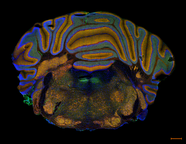
English: Immunofluorescent staining of the coronal section of the auditory brainstem with voltage-gated potassium channels Kv3.1 highlighted in green (anti-Kv3.1b + AlexaFluor 488, LED+Opal 520 filter), Kv3.3 in orange (anti-Kv3.3 + AlexaFluor 555, LED+Opal 570 filter) and nuclei in blue (DNA stain DAPI, LED+DAPI filter).
Kv3s ensure short action potentials by driving repolarisation back to rest. Kv3.1 and Kv3.3 channels can be found in many areas of the auditory pathway, as you can see from this brainscan, where the auditory brainstem is located below the cerebellum folds (a red string of cells are Purkinje neurons running between the granular and molecular layers of a cerebellum fold). We are focused on the Superior Olivary Complex in the auditory brainstem. Here, it is visible as two mirrored "chicken legs" at the bottom of the slice. We are specifically interested in the principal neurons of the Medial Nucleus of the Trapezoid Body or MNTB (being the inner – thinnest - part of the "chicken leg"). These cells host the largest synapses found in the brain, called Calyces of Held, often used as a model to study synaptic transmission and plasticity. We can use the elements of this pathway to study the ion channel's location in the cell body, axon, and synaptic terminal. Prep: frozen 12 µm sections from CBA WT mouse, P20. This image is a single optical section obtained using an Akoya Vectra Polaris slide scanner with a 20x objective. Scale bar: 0.5 mm.Українська: Імунофлуоресцентне зображення коронального зрiзу слухового стовбура мозку з потенціалзалежними калієвими каналами Kv3.1, виділеними зеленим кольором (анти-Kv3.1b + AlexaFluor 488, LED+Opal 520 фільтр), Kv3.3 помаранчевим кольором (анти-Kv3.3 + AlexaFluor 555, LED+Opal 570 фільтр) і ядрами синього кольору (барвник ДНК DAPI, LED+DAPI фільтр).
Родина iонних каналiв Kv3 забезпечує короткі потенціали дії, повертаючи реполяризацію до стану спокою. Канали Kv3.1 і Kv3.3 можна знайти в багатьох областях слухового шляху, як ви можете бачити на цьому скані частини мозку, де слуховий стовбур мозку розташований під складками мозочка (насичено-помаранчевий рядок клітин — це нейрони Пуркіньє, що проходять між зернистим і молекулярним шаром складок мозочка). Мої дослідження зосережденi на вищому оливковому комплексі (Superior Olivary Complex, SOC) в слуховому стовбурі мозку. Тут SOC виглядає як дві дзеркальні «курячі ніжки» внизу зрізу. Нас особливо цікавлять головні нейрони медіального ядра трапецієвидного тіла або MNTB (є внутрішньою – найтоншою – частиною «курячої ніжки»). Ці клітини містять найбільші синапси, знайдені в мозку, звані чашечками Хелда, які часто використовують як модель для вивчення синаптичної передачі та пластичності. Ми використовуємо елементи цього шляху для вивчення розташування іонних каналiв в тілі клітини, аксоні та синаптичному терміналі. Препарат: заморожений зріз (12 мкм) мозку миші CBA WT (дикий тип), P20. Це зображення є одним оптичним розрізом, отриманим за допомогою слайд-сканера Akoya Vectra Polaris з об’єктивом 20x. Масштаб: 0,5 мм.English: Kv3.1 and Kv3.3 channels in auditory brainstem of WT CBA mouse |
| Date | |
| Source | Own work |
| Author | Kseniia Bondarenko |
Licensing[edit]
- You are free:
- to share – to copy, distribute and transmit the work
- to remix – to adapt the work
- Under the following conditions:
- attribution – You must give appropriate credit, provide a link to the license, and indicate if changes were made. You may do so in any reasonable manner, but not in any way that suggests the licensor endorses you or your use.
| This image was uploaded as part of Science Photo Competition 2022 in Ukraine. |
File history
Click on a date/time to view the file as it appeared at that time.
| Date/Time | Thumbnail | Dimensions | User | Comment | |
|---|---|---|---|---|---|
| current | 02:06, 19 December 2022 |  | 3,396 × 2,623 (11.5 MB) | Morne Arin (talk | contribs) | Uploaded own work with UploadWizard |
You cannot overwrite this file.
File usage on Commons
The following 3 pages use this file:
File usage on other wikis
The following other wikis use this file:
- Usage on ua.wikimedia.org
- Usage on uk.wikipedia.org
Metadata
This file contains additional information such as Exif metadata which may have been added by the digital camera, scanner, or software program used to create or digitize it. If the file has been modified from its original state, some details such as the timestamp may not fully reflect those of the original file. The timestamp is only as accurate as the clock in the camera, and it may be completely wrong.
| Unique ID of original document | xmp.did:bf21f06e-a0c7-9746-8782-44713079cae8 |
|---|---|
| Software used | Adobe Photoshop 24.0 (Windows) |
| Date and time of digitizing | 20:45, 18 December 2022 |
| File change date and time | 01:07, 19 December 2022 |
| Date metadata was last modified | 01:07, 19 December 2022 |
| Horizontal resolution | 28.35 dpc |
| Vertical resolution | 28.35 dpc |
